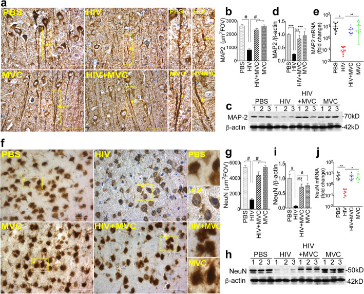
Fig. 5.
MVC prevented HIV-induced downregulation of the neuronal markers MAP 2 and NeuN. a: Immunohistochemistry analyses of MAP 2 expression in brain tissues (somatosensory cortex). b: Metamorph quantification of MAP 2 expression in all samples. MAP 2 levels in brain tissues were also quantified by Western blot (c) followed by densitometry quantification normalized to sample’s β-actin levels (d). f: immunohistochemistry analyses of NeuN expression in brain tissues (somatosensory cortex). g: Metamorph quantification of NeuN in all samples. NeuN levels in brain tissues were also quantified by Western blot (h) followed by densitometry quantification normalized to sample’s β-actin levels (i). MAP 2 (e) and NeuN (j) mRNA levels in brain tissues were quantified by real-time PCR. For panels a and f, images were at 40X. For panels b and g, 10 random FOV analyzed for each sample. The four animal groups included PBS, HIV, HIV + MVC, and MVC; 9 to 11 animals in each group. #P < 0.0001, ***P < 0.0003, **P < 0.004, *P < 0.015. Error bars represent SD

