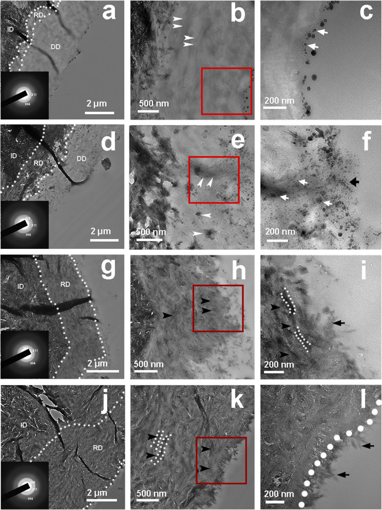
Fig. 6.
TEM images of dentin treated with the mineralizing film for 24 h (a–c), 48 h (d–f), 72 h (g–i) and 96 h (j–l). c, f, i and l are magnified images of b, e, h and k, respectively. At 24 h, spherical ACP nanoparticles were attached to the surface (c, white arrow). At 48 h, some nanoparticles (f, black arrow) were observed on the surface, and some were observed in the middle of the demineralized dentin layer (e, f, white arrow). The collagens became thicker and darker (e, white arrow). Rod-like crystals were detected on the surface of the remineralized dentin after 72 h (i, black arrow), and the demineralized dentin was fully mineralized and fused with the surface crystals of the dentin. A needle-like HAp layer was detected on the dentin surface at 96 h (l, black arrow). Both the black arrow and white dotted line (k, l) indicate the remineralized dentin collagen. ID, intact dentin; DD, demineralized dentin; RD, remineralized dentin

