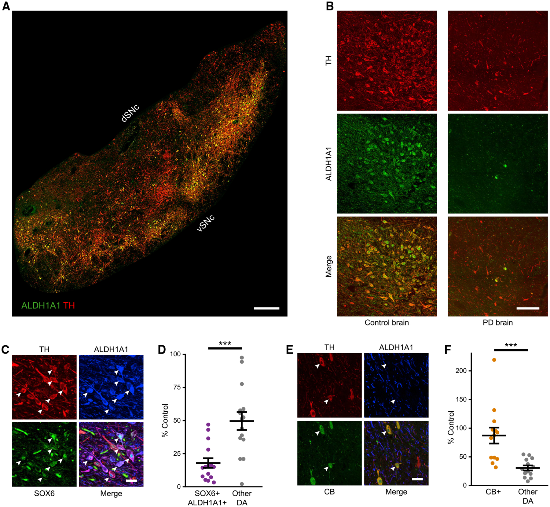
Figure 1. SOX6+,ALDH1A1+ neurons in human SNc are vulnerable in PD while CALBINDIN-D28k+ neurons are relatively spared.

(A and B) TH+,ALDH1A1+ labeling in control and PD brains. In PD brains, ALDH1A1 staining appears brighter in the nuclei compared to controls, as previously described in a 6-OHDA model (Stott and Barker, 2014).
(C) High magnification of SOX6+,ALDH1A1+ DA neurons (arrowheads) in a control brain.
(D) SOX6+,ALDH1A1+ DA neurons versus other DA neurons in hSNc of PD as a percentage of control brains (p = 3.1 × 10⁻⁴; controls are set at 100%, n = 14 controls, n = 15 PD).
(E) High magnification of CALBINDIN-D28k+ (CB+) DA neurons in a control brain.
(F) CB+ DA neurons versus other subtypes in PD as a percentage of control brains (p = 7.3 × 10⁻⁴, n = 14 controls, n = 15 PD).
Scale bars: (A) 1,000 μm, (B) 100 μm, and (C and E) 50 μm. Error bars are SEMs. vSNc, dSNc: ventral and dorsal substantia nigra pars compacta. hSNc: human substantia nigra pars compacta.
