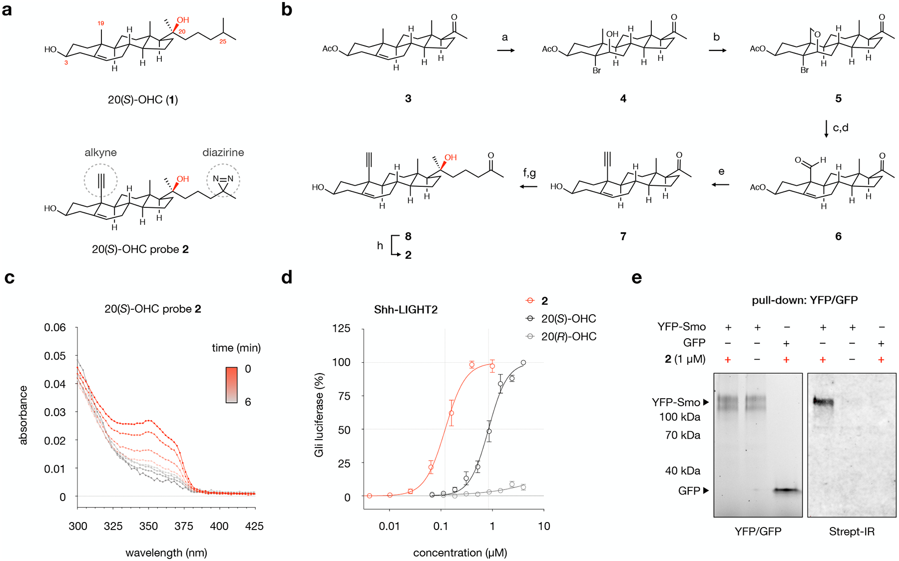
Fig. 1: Design, synthesis, and evaluation of 20(S)-OHC chemoproteomics probe 2.

a, Structures of 20(S)-hydroxycholesterol (20(S)-OHC, 1) and probe 2, which contains a diazirine at C25 for photocrosslinking and an alkyne at C19 for click chemistry.
b, Chemical synthesis of probe 2. Conditions: a. NBA, aq. HClO4, 1,4-dioxane, 0 → 23 °C, 77%; b. Pb(OAc)4, CaCO3, I2, hν, cyclohexane, 80 °C, > 99%; c. Zn, AcOH-H2O, 45 °C, 88%; d. PCC, Celite, CH2Cl2, 23 °C, 87%; e. (i) Seyferth-Gilbert reagent, t-BuOK, THF, −78 °C, 80%; (ii) Cs2CO3, MeOH, 23 °C, >99%; f. 2-(3-bromopropyl)-2-methyl-1,3-dioxolane, Mg, THF, 0 → 23 °C, 61%; g. HCl, THF, 23 °C, 94%; h. (i) NH3, MeOH, 0 °C; (ii) NH2HSO3, 0 → 23 °C; (iii) I2, Et3N, THF, 23 °C, 46%.
c, Irradiation of probe 2 (10 mM, DMF) with 368 nm light results in loss of diazirine absorption at 353 nm with a half-life of 1.4 minutes. Values are the average of triplicate measurements.
d, Treatment of Shh-LIGHT2 cells with 20(S)-OHC, 20(R)-OHC, or probe 2 demonstrates that 20(S)-OHC and 2, but not the unnatural epimer 20(R)-OHC, activate the Smoothened (Smo)-regulated Gli transcription factors. Values are the average of 3 biological replicates ± s.d.
e, Live HEK293T cells overexpressing YFP-Smo or GFP were treated with 2 or DMSO and irradiated, then YFP-Smo or GFP was isolated from membrane fractions using a GFP nanobody that recognizes both fluorescent proteins. Isolated proteins were subjected to a click reaction for biotinylation. Left: In-gel fluorescence analysis of membrane fractions from cells overexpressing YFP-Smo or GFP. Right: Detection of biotinylated proteins using Streptavidin IRDye 800CW (Strept-IR), demonstrating that 2-treated YFP-Smo-expressing cells are labeled with biotin. YFP-Smo-expressing cells treated with DMSO and GFP-expressing cells treated with 2 show no biotin labeling. The experiment was repeated three times independently with similar results.
