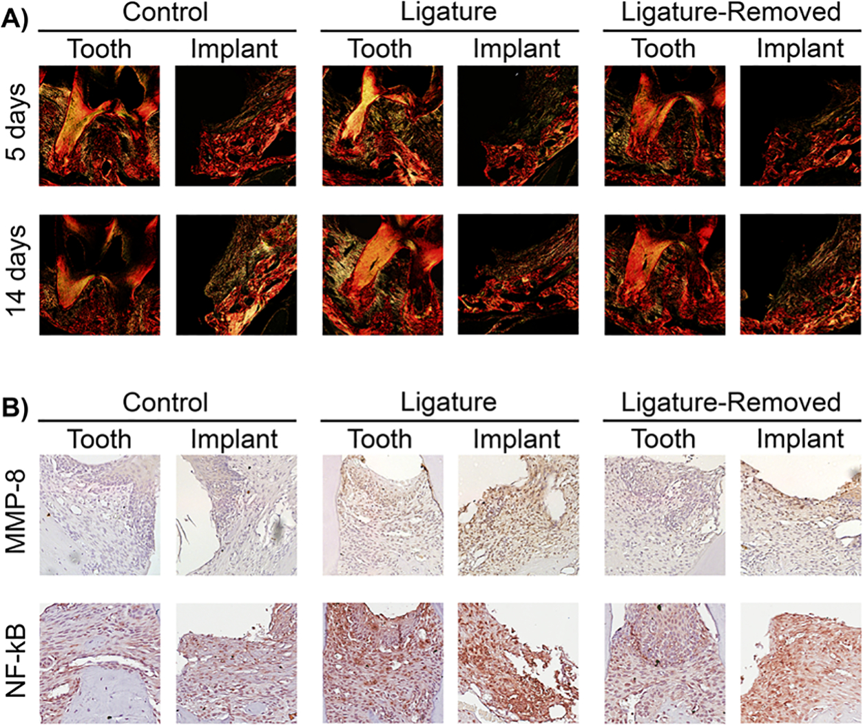
FIGURE 6.

Type 1 collagen, MMP-8, and NF-κB assessment in peri-implantitis and periodontitis after insult removal. A) Representative sagittal picrosirius red stained images of peri-implantitis and periodontitis control, ligature-retained, and ligature-removed groups 5 and 14 days after ligature removal at 10 × magnification under polarized light. Note greater type 1 collagen (yellow birefringence) destruction around implants than around teeth in the ligature-retained groups. New collagen fibers around implants are more apparent after 14 days of insult removal. B) The representative images of MMP-8 staining of peri-implantitis and periodontitis control, ligature-retained, and ligature-removed groups 5 days after ligature removal at 20 × magnification (top row). Note greater MMP-8 in the peri-implantitis ligature-retained and ligature-removed groups than the periodontitis groups. The representative images of NF-κB staining of peri-implantitis and periodontitis control, ligature-retained, and ligature-removed groups 5 days after ligature removal at 20 × magnification (bottom row). Note the greater NF-κB staining in the peri-implantitis ligature-retained and ligature-removed groups than the periodontitis groups
