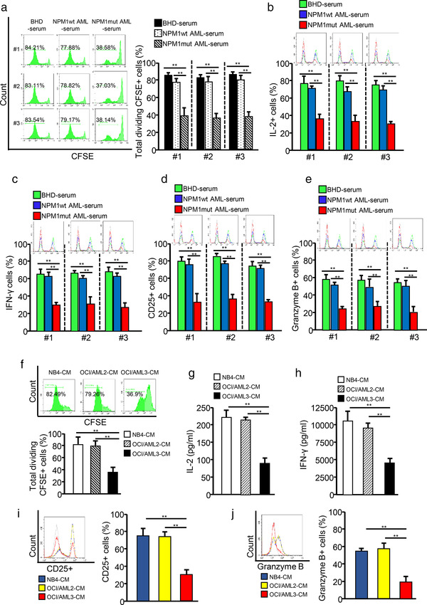
FIGURE 1.

NPM1mut AML cells impair CD8+ T cell function. CD8+ T cells were purified from blood samples from volunteers and stimulated with anti‐CD3 and anti‐CD28 antibodies to maintain their proliferative ability. (a) CD8+ T cells were pre‐labelled with CFSE and incubated with serum samples from patients with BHD, NPM1wt AML, or NPM1mut AML. CD8+ T cell proliferation was evaluated by FCM, and quantitative diagrams are shown. (b–e) CD8+ T cells were incubated with serum from patients with BHD, NPM1wt AML, and NPM1mut AML. Representative FCM analysis of IL‐2 (B), IFN‐γ (C), CD25 (d), and granzyme B (e) expression levels are presented. (f–j) CD8+ T cells were incubated with NB4‐CM, OCI/AML2‐CM, or OCI/AML3‐CM. (f) The proliferation of CD8+ T cells was evaluated by FCM. (g and h) IL‐2 (g) and IFN‐γ (h) levels in cell culture supernatants were analyzed for using ELISA kits. (i and j) The expression of CD25 (i) and granzyme B (j) was detected by FCM (*p < 0.05; **p < 0.01; ***p < 0.001; n.s, not significant)
