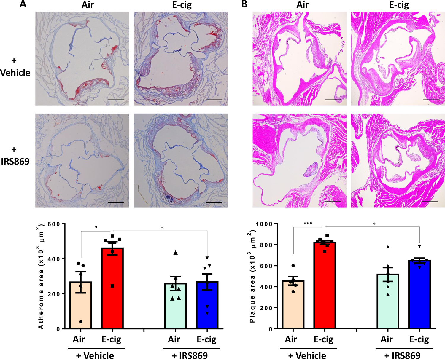
Figure 2. TLR9 inhibitor attenuated ECV-exacerbated atherosclerotic lesion development TLR9 inhibitor in ApoE−/− mice.

(A) Oil Red O (ORO) staining of the cross-sectioned aortic roots. Lipid deposition in plaques was quantified as ORO-positive lesion areas and expressed as percentage to the total area of the atheroma. (B). Cross-sectional hematoxylin-eosin staining to measure the intimal microscopic lesions in the aortic roots. Scale bar: 200 µm. Numbers of animals in each group: Air + Ctrl-ODN: n=5; E-cig + Ctrl-ODN: n=7; Air + IRS869: n=6; E-cig + IRS869: n=7. Data are shown as mean ± SEM and statistical multiple comparisons were made by one-way ANOVA with Tukey’s post hoc analysis. *P<0.05; ***P < 0.001.
