
Figure 1. MRI evidence of hypothalamic gliosis in association with body adiposity in children.
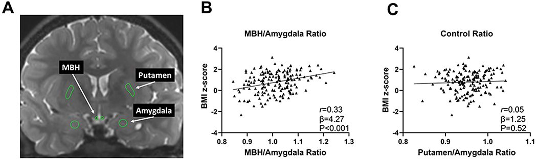
(A) Representative coronal slice of T2-weighted magnetic resonance imaging to assess T2 signal intensity from regions of interest in the MBH, amygdala, and putamen. Association of (B) MBH/amygdala and (C) putamen/amygdala T2 signal ratios with BMI z scores in children (n = 169). P values were calculated by linear regression and adjusted for age, sex, and study site. Pearson correlation coefficient was calculated for descriptive purposes. MBH, mediobasal hypothalamus