Fig. 2.

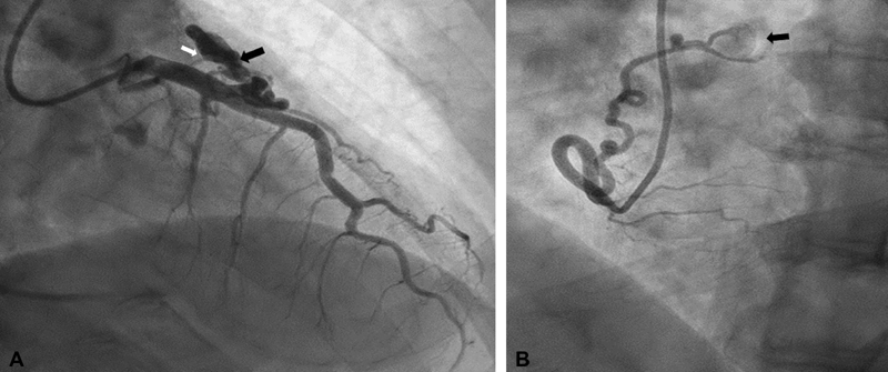
A 68-year-old male presented with inferior ST elevation myocardial infarction. He had coronary artery aneurysm with significant thrombus in right coronary artery (RCA) proximal. He had cover stent placed to proximal RCA. He was found to have two coronary artery fistulae. His stress test was negative. Cardiac computed tomography confirms the size and location of fistula. He was managed medically and did well in follow-up. ( A ) Right anterior oblique caudal view of left coronary artery showed aneurysmal dilatation of D2 (black arrow) along with small D2 to pulmonary artery fistula (white arrow). ( B ) Left anterior oblique view of RCA showed mid-size proximal RCA conus branch artery to pulmonary artery fistula (black arrow).
