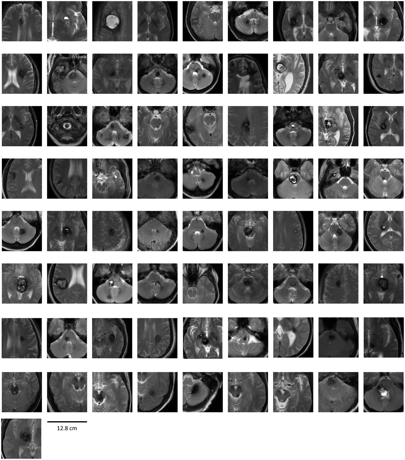
Figure 2. Magnetic resonance image thumbnails of 73 cases enrolled in CASH TR Project.

T2 images of CASH lesion in 73 consecutively enrolled FUBV cases through May 2020. Images obtained at enrollment, cropped to the region highlighting the CASH lesion at similar scale (bar = 12.8 cm). A diagnostic CASH event must have occurred within the prior 12 months (images of diagnostic hemorrhage not shown).
