
Figure 7.
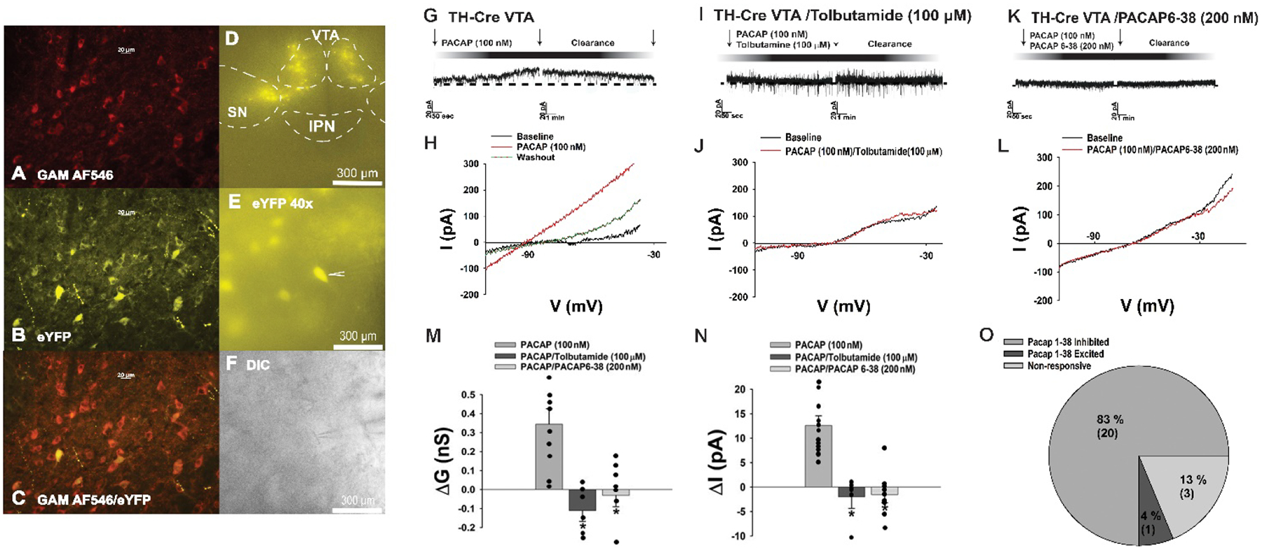
PACAP inhibits A10 dopamine neurons in slices from male TH-cre mice via a PAC1 receptor-mediated activation of KATP channels. A, TH immunostaining in the VTA. B, eYFP signal seen in these A10 dopamine neurons. C composite overlay. D & E, eYFP signal from A10 dopamine neurons in VTA slices seen at 4X and 40X. F, DIC image of the recorded A10 dopamine neuron seen in E. G-O, PACAP (100 nM; n = 12) produces a robust and reversible outward current in A10 dopamine neurons that is associated with an increased K+ conductance and abrogated by PACAP6–38 (n = 9) and tolbutamide (n = 6). Arrows indicate where I/Vs were conducted. Bars represent means and lines 1 SEM of the change in membrane current (M; pA) membrane slope conductance (M; nS) and current (N; pA). *p < 0.05 relative to PACAP alone, one-way ANOVA/LSD.