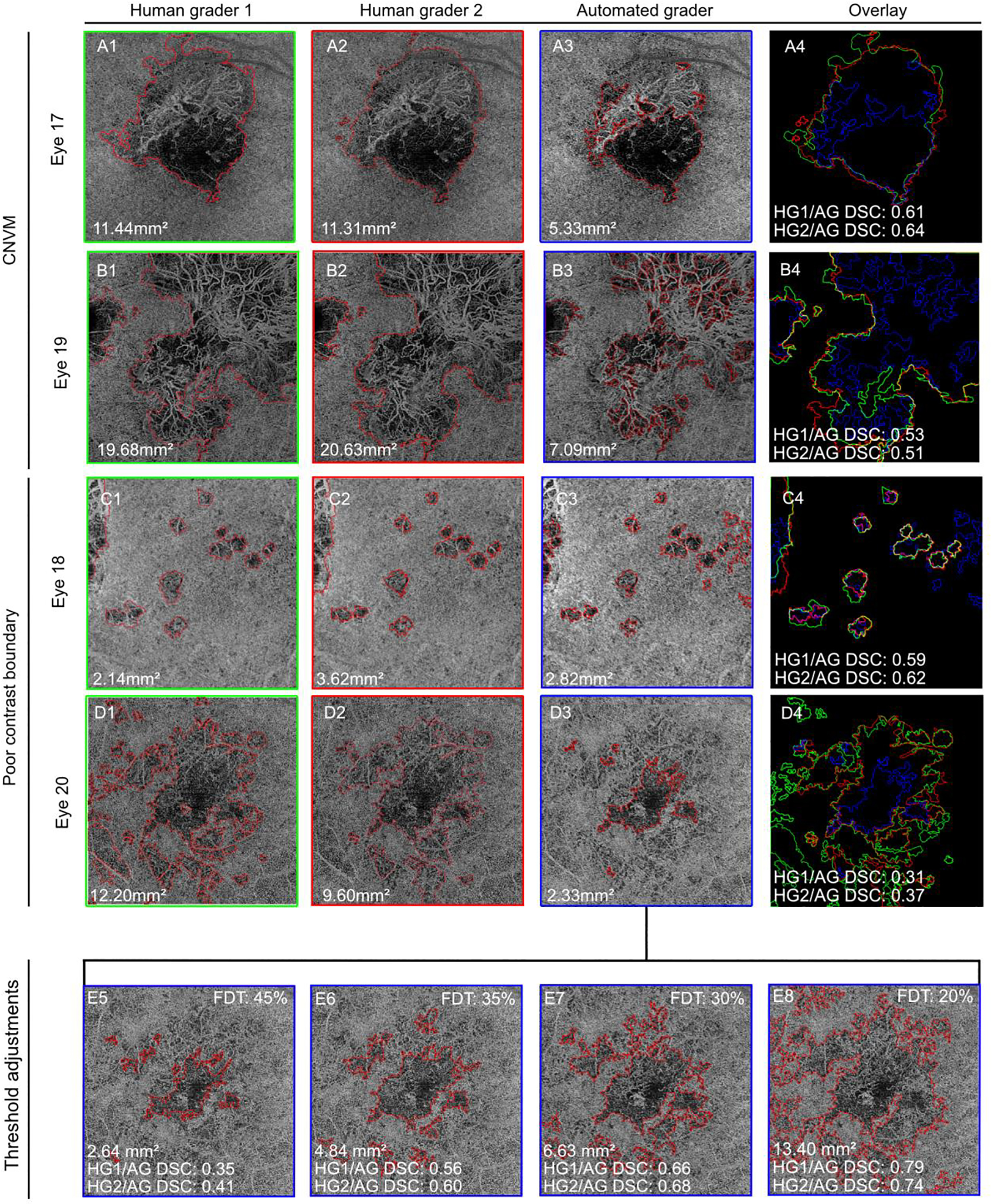
FIGURE 3. Poor inter-grader agreement is associated with the presence of CNVM and poor contrast lesion boundaries:

(A) Eye 17, from a patient with multifocal choroiditis. (B) Eye 19, from a patient with birdshot chorioretinopathy. When visible in the choriocapillaris slab, the choroidal neovascular membrane (CNVM) is included in the lesion area by the human graders (HGs) but excluded by the algorithm (AG). (C) Eye 18, from a patient with multifocal choroiditis. Total area is similar, but exact location of the lesion boundaries varied between graders. (D) Eye 20, patient with acute multifocal pigmented placoid epitheliopathy. The AG identified a significantly smaller lesion than both human graders. (E) Alternative AG lesion boundaries generated by decreasing the flow deficit density threshold (FDT). This change allowed the AG to include more subtle FD abnormalities into the lesion area, and improved agreement with human graders.
CNVM = choroidal neovascular membrane, FDT = flow deficit density threshold, HG1 = human grader 1, HG2 = human grader 2, AG = algorithm
