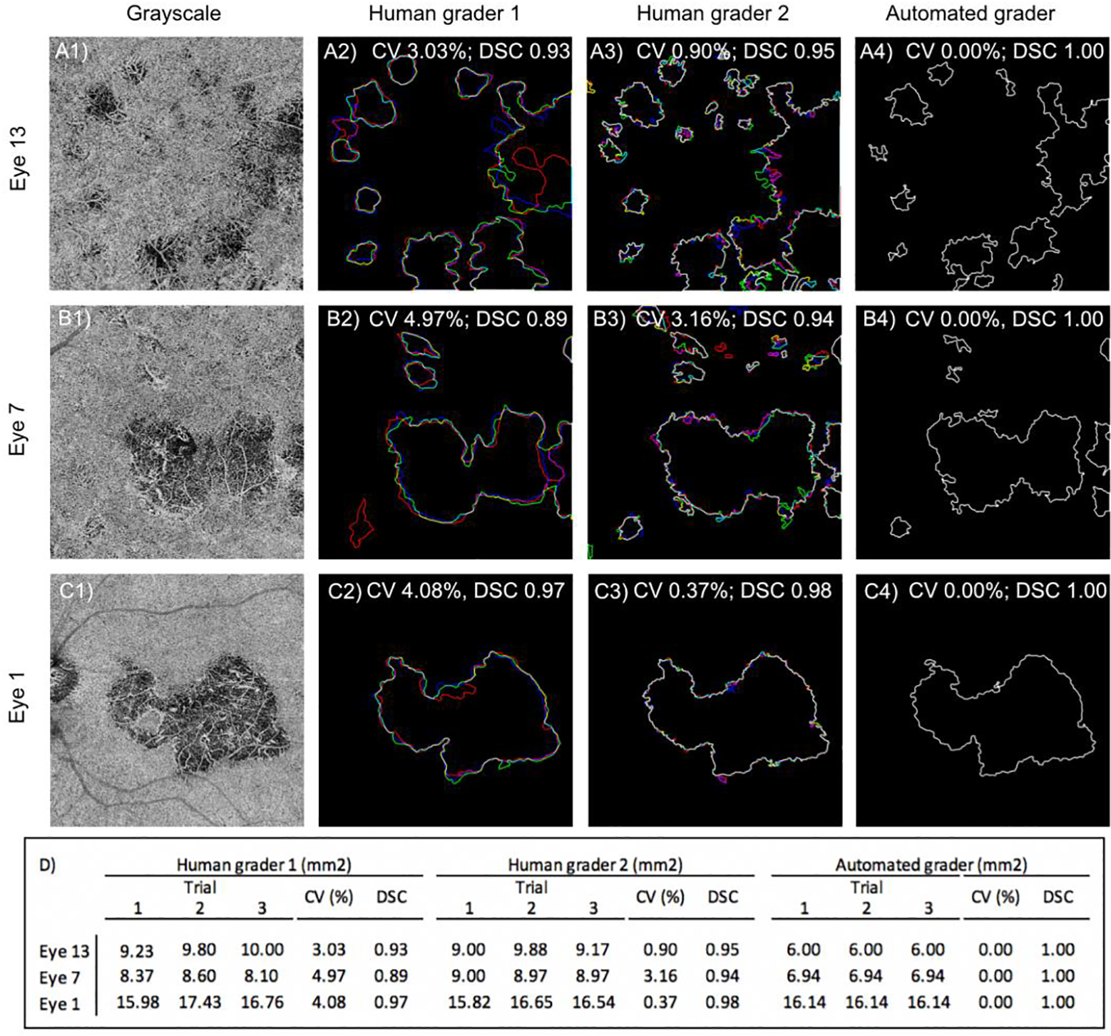
FIGURE 4. The algorithm performs better on repeat segmentation of the same image than human graders:

(A) Eye 13, from a patient with birdshot chorioretinopathy. (B) Eye 7, from a patient with relentless placoid chorioretinopathy. (C) Eye 1, from a patient with serpiginous choroiditis. The first column shows the CC en face image that was segmented by each grader. Each column to the right shows the results for HG1, HG2, and the algorithm (AG). For each image, the graders segmented the lesion three times: trial 1 (red), trial 2 (green), trial 3 (blue). Areas of perfect overlap are shown in white. (D) Table showing the total lesion area obtained by each grader at each independent scoring session, coefficient of variance (CV), and average DSC.
