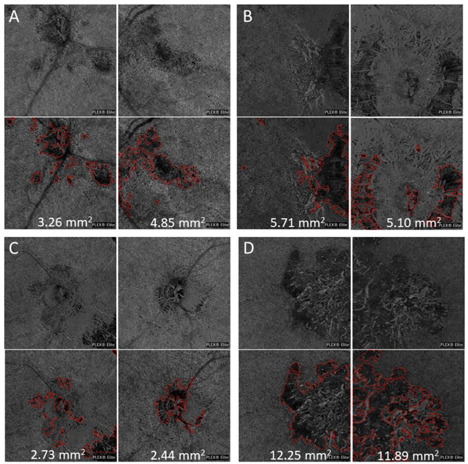
Figure 7: Application of automated lesion segmentation and area quantification to images obtained from a second clinical location.

The choriocapillaris (CC) slab from the right and left eyes from four patients with posterior uveitis (top row in each panel) were analyzed and the CC flow deficits were outlined (red) and lesion area quantified in mm2 (bottom row in each panel). (A) Chorioretinitis with secondary peripapillary CNVM associated with undifferentiated connective tissue disease with concomitant small vessel vasculitis (B) Multifocal choroiditis with secondary CNVM (C) Multifocal choroiditis (D) Serpiginous choroiditis.
