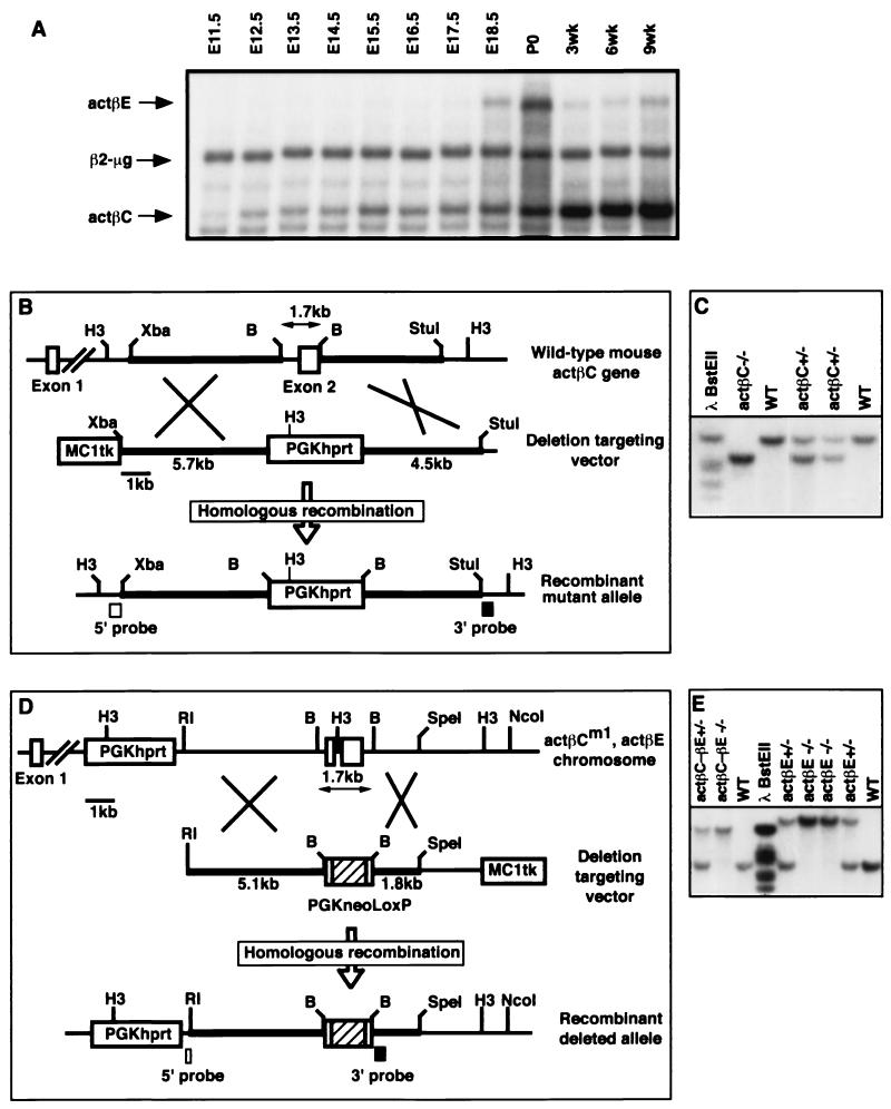
FIG. 1.
Expression of activin βC and activin βE in the livers of embryos and adult mice and gene targeting constructs for activins βC and βE. (A) Expression of actβC and actβE in mouse embryonic and adult livers was detected using RNase protection assays. β2-Microglobulin was used as the internal control. Five micrograms of total RNA from C57BL/6 mice was hybridized to probes specific for activin βC, activin βE, and β2-microglobulin. Shown is a representative autoradiograph repeated in four independent experiments. (B) The targeting strategy used to delete exon 2 of the mouse activin βC gene. The construct contains an MC1tk expression cassette for negative selection and a PGKhprt expression cassette for positive selection. Homologous recombination will delete all of the coding sequence in exon 2. (C) Southern blot analysis of tail DNA from F2 mice at weaning. The 3′ probe identifies the wild-type 13.8-kb band and the 8.3-kb mutant band. (D) The targeting construct to delete all of the coding exons of the actβE gene. Homologous recombination within the homology arms will replace the entire coding region of activin βE with a floxed (vertical-box-flanked) PGKneo-positive selectable marker cassette. The MC1tk cassette is used for negative selection. Due to the close physical proximity of activin βC and βE loci on the same chromosome, ES cells carrying the actβCm1 mutation (cell line βC45-F1, which was used previously for germ line transmission of actβCm1) were electroporated. (E) Southern blot analysis showing weaned F2 mice homozygous for the actβEm1 mutation (19.2-kb band) or the double mutant allele (actβCm1-actβEm1; 13.8-kb band). The wild-type band migrates at 5.3 kb. H3, HindIII; B, BamHI; Xba, XbaI; RI, EcoRI; WT, wild type.

