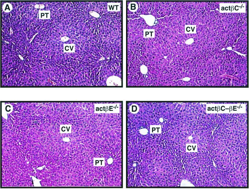
FIG. 3.
Histological analysis of livers from 42-day-old wild-type and null mice. Liver tissue from C57BL/6-129/SvEv hybrid mice was formalin fixed and stained with hematoxylin and eosin. The liver histology of the homozygous mutant mice compared to that of wild-type mice showed the expected liver lobular organization of the hepatic parenchyma. All sections were photographed at a magnification of ×100. (A) Wild-type mouse (WT); (B) actβCm1/actβCm1 mouse (actβC−/−); (C) actβEm1/actβEm1 mouse (actβE−/−); (D) actβCm1-actβEm1 (actβC-βE−/−) homozygous mutant mouse. CV, central vein; PT, portal triad.

