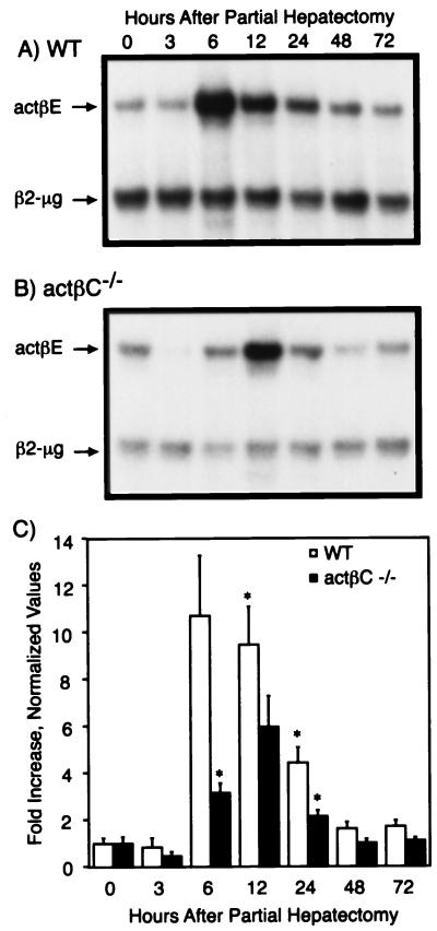
FIG. 6.
Expression of actβE before and after partial hepatectomy. (A and B) Remnant or whole livers from wild-type (WT) (A) and actβCm1/actβCm1 (actβC−/−) (B) mice were collected at 0, 3, 6, 12, 24, 48, and 72 h after partial hepatectomy. Representative autoradiographs are shown. Expression of actβE in the livers was assayed by RNase protection with β2-microglobin (β2-μg) as the internal control. (C) The protected bands were quantitated and visualized as fold increase over the 0-h point ± standard error of the mean for each genotype. An asterisk denotes time points where expression of actβE in activin βC knockout or wild-type mice was statistically significantly different from the time zero value for the same genotype (P < 0.05) by Student's t test. The numbers of mice analyzed at each time point were three to four for wild type and actβCm1/actβCm1.

