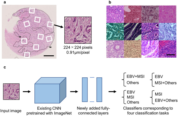
Figure 1.
Image processing and the architecture of neural networks. (a) Representative image of tissue microarray prepared from cases of gastric cancer. Many small images (224 × 224 pixels) were sampled from annotated tumor areas at random positions and angles. Scale bar: 500 µm. (b) Sampled images after data augmentation (random change of color tone and blurring). Scale bar: 100 µm. (c) The architecture of neural networks: Fine-tuning of existing CNNs (VGG16, VGG19, ResNet50 and EfficientNetB0) was adopted. Classifiers corresponding to four classification tasks were added on the top. CNN—convolutional neural networks.

