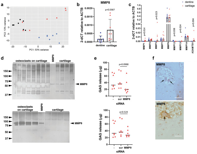
Figure 4.
MMP8 and MMP9 drive osteoclast-mediated release of GAG from cartilage. (a) Principal Component Analysis (PCA) plot of osteoclasts on acellular cartilage (red), dentine (blue) and cell culture plastic (black), n = 7. (b,c) RT-qPCR analysis of differences in expression of MMPs by osteoclasts differentiated on dentine or acellular cartilage: (b) MMP8 (n = 8; T test); (c) MMP1, 2, 3, 7, 9, 12, 13, 14 and ADAMTS1 (n = 8; multiple Mann–Whitney). (d) Representative zymography of MMP8 and MMP9 from the conditioned media of osteoclasts differentiated on acellular cartilage compared to recombinant protein and cartilage alone. (e) GAG released by osteoclasts differentiated on acellular cartilage and transfected with siRNA targeting MMP8 or MMP9 on day 7 of differentiation, versus no siRNA (−) and scrambled siRNA (scr) controls (n = 7; Kruskal–Wallis ANOVA). (f) Human OA tissue sections stained for MMP8 (top) and MMP9 (bottom). Arrows indicate multinucleated osteoclasts. Scale bar = 100 µm.

