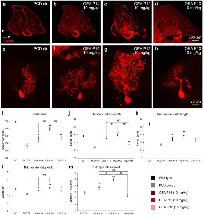
Fig. 4.
In vivo effect of different acute administration of OEA on the morphology and survival of the Purkinje cells of the PCD mouse analyzed at P30 (second set of experiments). (a–d) Micrographs of PCD cerebellar vermis slices labeled with calbindin (Cb28k, red) at different OEA acute administrations at a dose of 10 mg/kg: control (a), at P14 (b), at P12 (c), and at P10 (d); an increase in the Purkinje cell density can be qualitatively observed when OEA is administered at P12. (e–h) Micrographs of Purkinje cells labeled with calbindin (Cb28k; red) in PCD animals administered with OEA (10 mg/kg) at P14 (f), P12 (g), and P10 (h); a notable improvement in the Purkinje cell arborization can be qualitatively observed when OEA is administered at P12. (i–l) Quantification of the effect of OEA on Purkinje cells morphology; note that the neuroprotective effect of OEA administered acutely follows an inverted U-shaped time-response curve, with acute administration of OEA at P12 being the most effective treatment for stabilizing Purkinje cell morphology, as the morphological parameters reached values similar to those of WT animals. (m) Quantification of the effect of OEA on Purkinje cell survival. Data are represented as mean ± SEM; n = 7 each experimental group; one-way ANOVA followed by Bonferroni’s post hoc test for (i–m); *p < 0.05, **p < 0.01 for differences between experimental group and control group (PCD without treatment); #p < 0.05, ##p < 0.01 for differences between the different OEA treatments. Data from WT animals has been used only as a reference, not to compare

