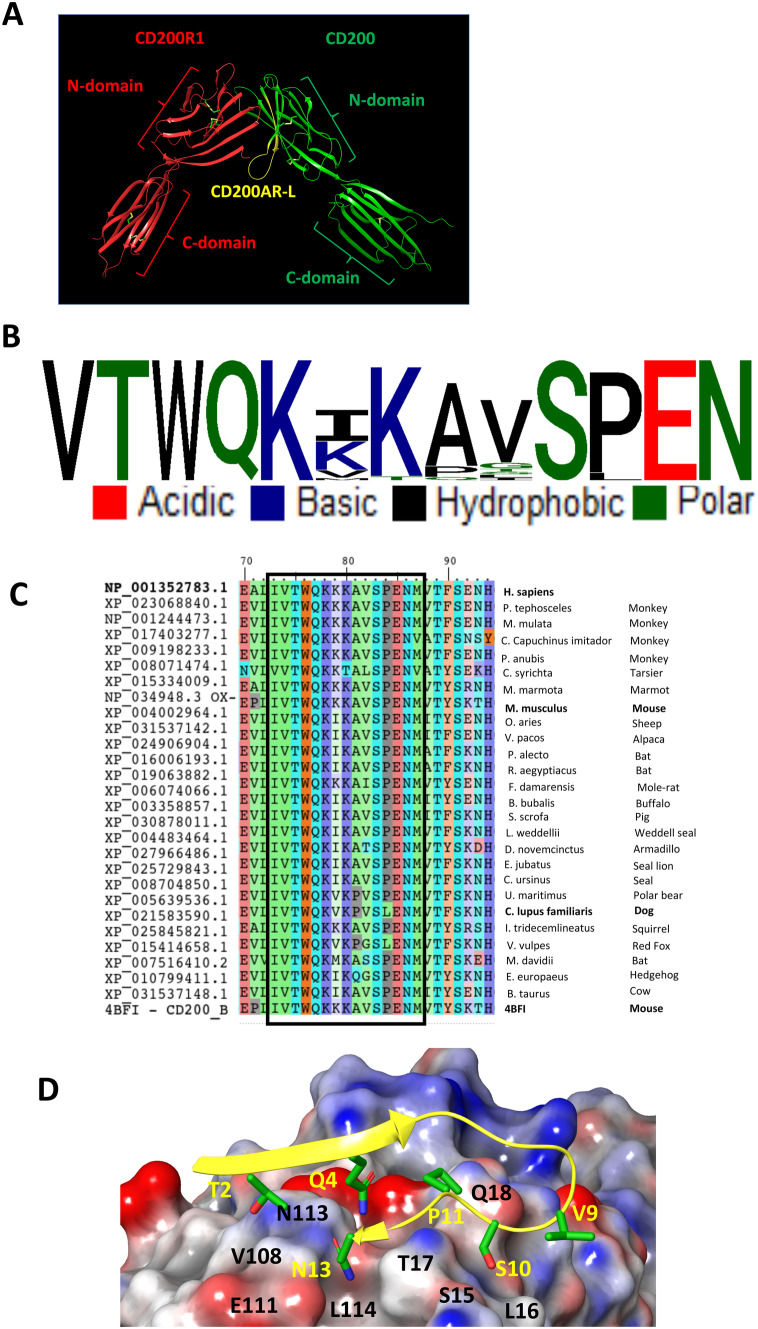
Fig. 1.
CD200AR-L modeling. A Cartoon representation of the mouse CD200R1 (red) and CD200 (green) crystal structure (PDB: 4BFI) with the highlighted sequence location of CD200AR-L (yellow). Association between CD200 and CD200R1 involves the 2 N-domains which includes CD200AR-L residues 32–44 region that accounts for 42% of the CD200R1 binding site. B Sequence conservation of CD200AR-L across the 26 available mammalian CD200 sequences from NCBI. C Multiple sequence alignment of mammalian CD200 sequences from NCBI. The highly conserved CD200AR-L sequence motif (boxed) is highlighted. D Electrostatic potential surface of the binding site highlighting the electrostatic complementarity between CD200AR-L and CD200R4 residues

