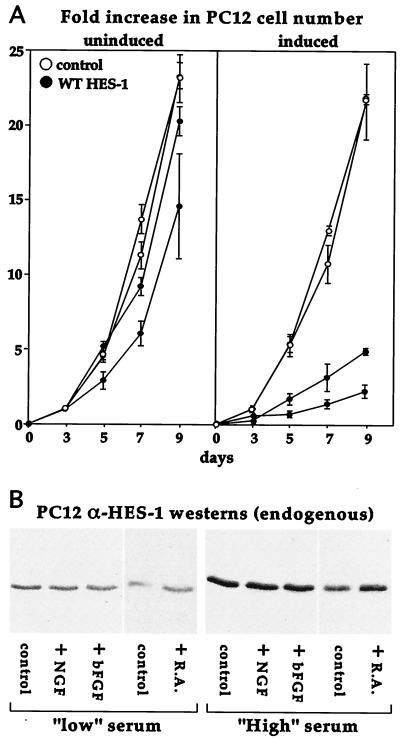
FIG. 3.
Induction of WT HES-1 inhibits PC12 cell proliferation. (A) Two lines of control and two lines of WT HES-1 PC12 cells were maintained either with or without tetracycline for 3 days, then equal numbers of cells were passaged in triplicate to measure proliferation. The cells were maintained up to 9 additional days (with or without tetracycline), and a set of plates for each cell type was counted at the 3-, 5-, 7-, and 9-day time points. The experiment was repeated, and the data for both experiments is shown in the graph as the average fold-increase in cell number with the error as the standard deviation (A). The growth of uninduced WT HES-1 cells was slightly lower than control lines, perhaps due to leakage of the exogenous HES-1 (A, left panel). In contrast, the growth of the WT HES-1 cells was markedly lower upon induction of HES-1 (A, right panel). Induction of EGFP did not greatly affect the growth of control cells. The rate of proliferation of the WT HES-1 cells may be overestimated in the induced panel, since prolonged induction could select for the lower-inducing, faster-growing cells. (B) Western analysis of endogenous HES-1 in parental PC12 cells showed that HES-1 was induced by serum in the media (B, compare the panels of low-serum-level-maintained cells to the panels of normal (high)-serum-level-maintained cells). Addition of the differentiation agents NGF or basic FGF did not appear to affect HES-1 levels in either serum condition. In contrast, retinoic acid (R.A.), which halts PC12 cell proliferation but does not differentiate the cells, did raise HES-1 protein levels slightly.

