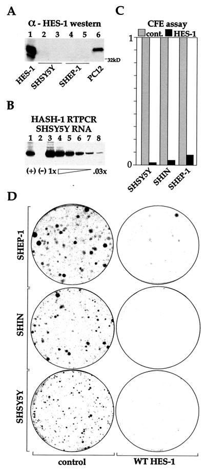
FIG. 6.
WT HES-1 inhibits proliferation in neuroblastoma cell lines. Three neuroblastoma cell lines (SHSY5Y, SHEP1, and SHIN) were tested for CFE following transfection with WT HES-1. These cell lines do not have significant expression of HES-1 protein, as shown by Western analysis of two of the lines in panel A. Transiently expressed HES-1 (lane 1) and endogenous PC12 cell (lane 6) lysates were run as positive controls for the detection of HES-1 protein. Lanes 3 and 5 are from lysates of NGF-treated cells. As might be anticipated by the lack of HES-1, expression of the MASH-1 gene can be detected in these cells, shown by reverse transcription-PCR of the SHSY5Y line in panel B. The (+) lane (lane 1) is a cDNA-positive control, the (−) lane (lane 2) is a reverse-transcriptase-negative control, and lanes 3 through 8 are serial dilutions of the input reverse transcription reaction for PCR amplification. MASH-1 mRNA has also been detected by reverse transcription-PCR in the SHEP and SHIN cell lines, as well as in LAI 5S, LAI 55N, and BEI YC neuroblastoma cell lines (data not shown). The greatly reduced colony formation of the HES-1-transfected cell lines is shown in panel C, and representative plates with Coomassie blue-stained cell colonies are shown in panel D. The near absence of colonies expressing WT HES shows that the inhibition of cell proliferation is a general property of WT HES-1 overexpression.

