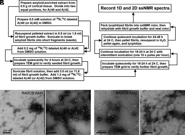
Fig. 1.
Preparation of brain-seeded Aβ fibrils for solid-state NMR (ssNMR). (A) Flowchart representation of the protocol for preparation of isotopically labeled Aβ40 and Aβ42 fibril samples by seeded growth from amyloid in human brain extract. (B) TEM images of negatively stained Aβ40 fibrils prepared from frontal lobe tissue of RADC subject 5. Images are shown after the initial 4-h incubation step (Left) and after the subsequent 18- to 24-h incubation step (Right). (C) Same as in A but for Aβ42 fibrils. (Scale bars: 200 nm.)

