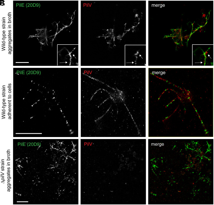
Fig. 2.
PilV is distributed throughout the N. meningitidis T4P. (A–C) dSTORM images of N. meningitidis grown in liquid broth (WT or ΔpilV) or attached to endothelial cells (WT). Images were acquired on a Leica SR GSD 3D system. A total of 50,000 frames were recorded and reconstructed using LAS X Software. (Scale bar, 2 μm.)

