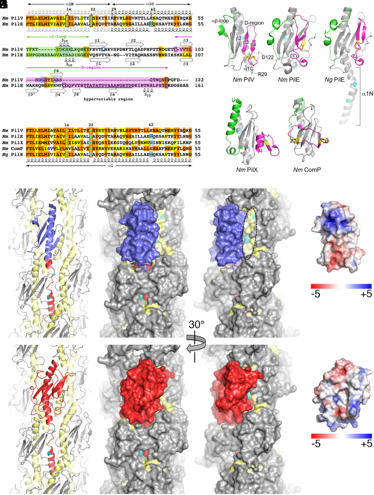
Fig. 3.
X-ray crystal structure of N. meningitidis PilV and model of PilV within N. meningitidis T4P. (A) Sequence alignment of the N. meningitidis (Nm) minor pilin PilV (National Center for Biotechnology Information WP_002244869) and the major pilin PilE (WP_014573675). Identical residues are highlighted orange and conserved residues in yellow. Helix-breaking Gly and Pro in α1 are highlighted in cyan and are boxed in black, as are Cys. Residues implicated in host-cell adhesion are boxed in blue. Secondary structures are indicated. The hypervariable region of PilE is underlined. (B) Crystal structure of N. meningitidis recombinant PilV, residues 29 through 122. The αβ-loop between α1 and the β-sheet is colored green, and the D-region, delineated by the disulfide-bonded cysteines, is magenta. Cysteines are shown as yellow sticks. The histidine at position 42 is colored cyan. (C) N. meningitidis major pilin PilE (residues 29 through 161, Protein Data Bank [PDB] 5JW8). (D) N. gonorrhoeae (Ng) major pilin PilE (full-length, residues 1 through 158, 2HI2). (E) Sequence alignment of α1 for N. meningitidis pilins and N. gonorrhoeae PilE, colored as in A (Nm PilX, CWT82783; Nm ComP, WP_002218144; Ng PilE, P02974). (F) N. meningitidis minor pilin PilX (residues 28 through 147, 2OPE). (G) N. meningitidis minor pilin ComP (residues 29 through 118, 5HZ7). Residues at positions 14 and 22 (N. gonorrhoeae PilE) and 42 (all pilins) are colored cyan. (H) The globular domain of a PilE subunit in the N. meningitidis T4P reconstruction (5KUA) was replaced with the rPilV structure (blue) by superimposing the N- and C-terminal ends of α1C, leaving only α1N of PilE (red). All other PilE subunits are colored gray, with α1 (residues 1 through 55) shown in yellow and α1N residues Gly14 and Pro22 in cyan. The model is shown in cartoon (Left) and space-filling representations (Middle and Right). The filament has been rotated about its long axis in the right panel to show how the narrower PilV globular domain exposes α1N of a higher PilE subunit (dashed oval). (I) For comparison, the N. meningitidis T4P reconstruction is shown with a single PilE subunit colored red. (J, K) Electrostatic surface representation of PilV (J) and PilE (K), shown in approximately the same orientation as in H and I.

