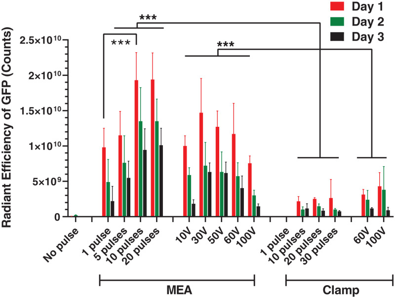
Fig. 4.
GFP expression in rat skin after electroporation. Radiant efficiency of GFP fluorescence in the skin on different days after delivery of GFP reporter plasmid by electroporation using an ePatch giving 1 to 20 pulses of ∼300 V with a waveform like that shown in Fig. 2B or using a conventional exponential decay electroporation pulser at controlled peak voltage (10 V to 100 V) with decay time constants (τ = 49 ms to 57 ms). Pulses were applied using an MEA or a clamp electrode. Data represent mean ± SD (n = 5 or 6) (***P < 0.001).

