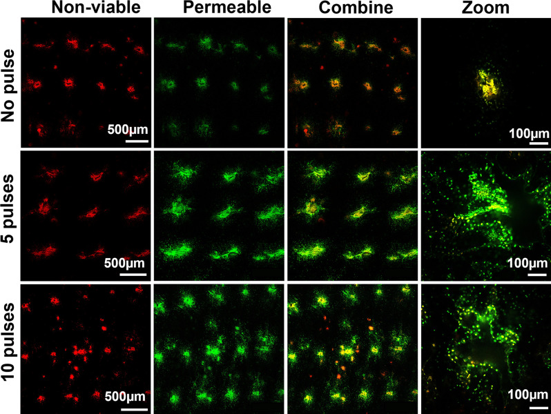
Fig. 5.
Cell membrane permeabilization and cell viability in mouse skin in vivo after electroporation by ePatch. Representative images show nonviable cells (red color) and cells with permeabilized membrane (green color) in the skin after electroporation with 0, 5, or 10 pulses by ePatch. Nonviable cells were identified in the mouse skin after insertion of MEA without electroporation (no pulse), suggesting that a small number of cells were damaged by microneedle electrode insertion alone. The red and green signals are colocalized in the insertion holes because nonviable cells are also permeable to the SYTOX Green. After 5 and 10 pulses, the red signal did not increase in the skin, while the green signal became more dispersed in the skin, suggesting transient cell permeability induced by the piezoelectric pulses with little effect on cell viability.

