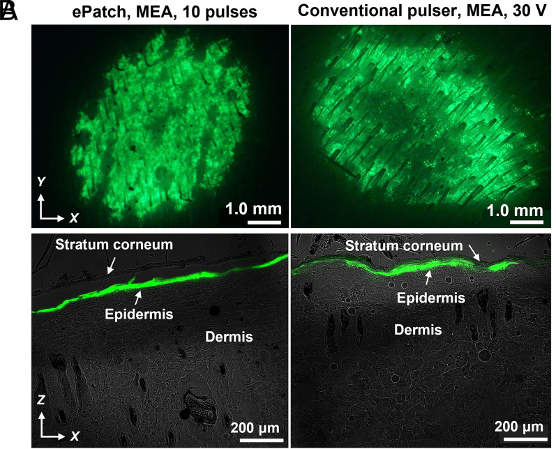
Fig. 6.
Fluorescence micrographs of rat skin imaged 1 d after delivery of GFP reporter plasmid by electroporation. Representative images are shown after electroporation using an MEA with 10 piezoelectric microsecond pulses administered by ePatch (Left) and with a single, exponential decay millisecond-long pulse (30 V, τ = 54 ms) administered by a conventional electroporation pulser (Right). After electroporation in vivo, skin was biopsied and imaged by (A) stereo fluorescence microscope on the skin surface and (B) laser scanning confocal microscopy as cryosections of the skin. The green color indicates GFP fluorescence. Skin anatomy is indicated in B.

