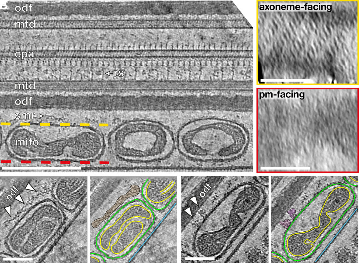
Fig. 2.
Ordered protein arrays on the OMM interact with the cytoskeleton. (A) Slice through a cryotomogram of an FIB-milled, horse sperm midpiece showing mitochondria (mito), the submitochondrial reticulum (smr) ODFs (odf), microtubule doublets (mtd), and the central pair apparatus (cpa). Note how individual complexes (like the radial spoke, rs) are visible in the raw tomogram. The ordered protein array is only found on the axoneme-facing surface (yellow) of midpiece mitochondria and not on the plasma membrane–facing surface (red). (B and C) Slices through a cryotomogram of an FIB-milled, horse sperm midpiece showing how the array directly interacts with the submitochondrial reticulum to anchor mitochondria to the flagellar cytoskeleton (arrowheads). In Right panels, the OMM is traced in green, the inner mitochondrial membrane in yellow, and the plasma membrane in blue. (Scale bars: [A] Left, 250 nm, Insets, 100 nm, and [B and C] 100 nm.)

