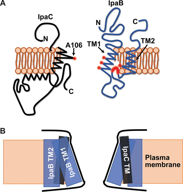
FIG 6.
Model of IpaB and IpaC in membrane-embedded translocons. (A) Schematic of IpaB (blue) and IpaC (black) in the plasma membrane (orange). Residues in the transmembrane domains that are extracellularly accessible to PEG5000-maleimide are highlighted in red. Although only one molecule of each translocase is shown, the pore is hetero-oligomeric. Images are not drawn to scale. (B) Schematic of the shape of the channel of the S. flexneri translocon pore, formed by the transmembrane domains of IpaB and IpaC. IpaC TM (black rectangle), IpaB TM1 and TM2 (blue rectangles), plasma membrane (orange box) are shown. The proposed shape of the pore channel is outlined (black lines).

