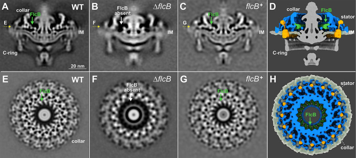
FIG 2.
ΔflcB (Δbb0058) mutant cells show defects in the flagellar collar structure. (A to C) A central section of the subtomogram averages (16-fold symmetrized) of the WT, ΔflcB, and flcB+ flagellar motors, respectively. The middle portion of the collar is absent in the ΔflcB motor. (E to G) A top view corresponding to the motor structures shown in panels A to C (indicated by yellow arrows), respectively. (D and H) A cross and top view of the 3D rendering of the WT flagellar motor, respectively. The FlcB protein is shown in green. Only the collar, stator, and inner membrane (IM) are shown in panel H.

