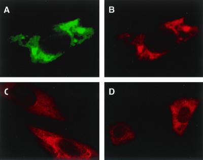
FIG. 1.
Immunofluorescence analysis of cultured cells on the intracellular distribution of GTPBP1. COS-7 cells were transfected with an HA-tagged mouse GTPBP-1 expression vector, pSRHAGP1, and cultured on coverslips. Forty-eight hours after the transfection, cells were double stained with anti-HA monoclonal antibody (12CA5) and anti-GTPBP1 polyclonal antibody (GP1a). Staining signals with 12CA5 were visualized by FITC-labeled anti-mouse IgG (A), and the GP1a staining signal was visualized by Cy3-labeled anti-rabbit IgG (B). A rat aortic smooth muscle cell line, A10 (C), and mouse peritoneal macrophages (D) were also cultured on coverslips and stained with GP1a. Magnifications, ×850 (A, B, and C) and ×340 (D).

