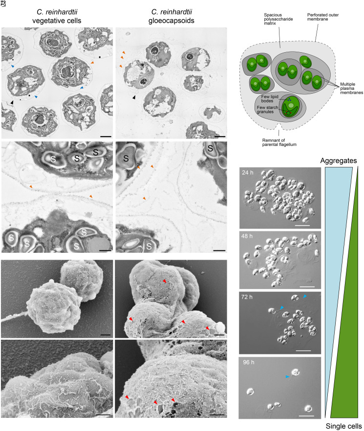
Fig. 2.
Comparison of the ultrastructure of C. reinhardtii vegetative cells with gloeocapsoids and disassembly of gloeocapsoids after stress relief. (A) Transmission electron microscopy pictures of vegetative C. reinhardtii cells (Left) and gloeocapsoids (Right). Vegetative cells are surrounded by a single membrane (orange arrows) and contain multiple starch granules (“S”; 26.2 ± 7.9 per cell; n = 5). Gloeocapsoids are surrounded by up to three membranes (orange arrows) and a polysaccharide matrix. In the individual cells, there are fewer starch granules (“S”; 6.2 ± 2.9 per cell; n = 5). In their periphery, vegetative cells show secretory vesicles (blue arrows), which are missing in cells embedded in gloeocapsoids. (Scale bars: 2 μm [Top] and 500 nm [Bottom]). (B) Scanning electron microscopy pictures of C. reinhardtii vegetative cells (Left) and gloeocapsoids (Right). The outer membrane of vegetative cells is intact. In gloeocapsoids, the outer membrane exhibits a perforated net-like structure (holes are marked with red arrows). (Scale bars: 1 μm [Top] and 500 nm [Bottom]). (C) Schematic model of the structure of a gloeocapsoid. (D) Disassembly of gloeocapsoids after removal of azalomycin F. Flagellated single cells dominated the culture 96 h after azalomycin F was removed. Visible flagella are indicated with blue arrows. (Scale bars: 20 μm.)

