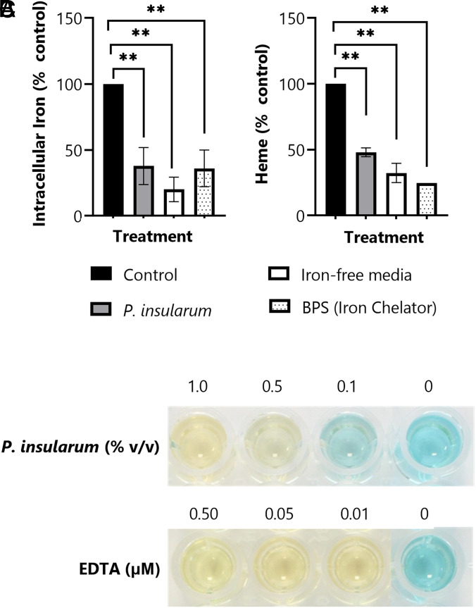
Fig. 3.
P. insularum treatment impacts iron homeostasis. (A) Intracellular iron levels in WT cells grown in control media or treatment media containing 0.05% v/v P. insularum homogenate, iron-free media, or 0.1 µM BPS iron chelator. Cells were grown to midlog, lysed, and intracellular iron was quantified using ICP-MS. Results shown are average and SD from three independent experiments with three technical replicates each. **P < 0.01; Student’s t test comparison to control media. (B) Heme levels in WT cells grown in the same conditions as panel A. Protein was extracted and heme levels were measured using the triton methanol method via a standard curve with known concentrations of hemin and normalization to control levels. Results presented are the average and SD calculated from three independent experiments with three technical replicates each. **P < 0.01; Student’s t test comparison to control media. (C) Iron chelation activity of the P. insularum homogenate compared to EDTA iron chelation was measured in a cell-free CAS assay. A color change from blue to yellow is indicative of iron chelation.

