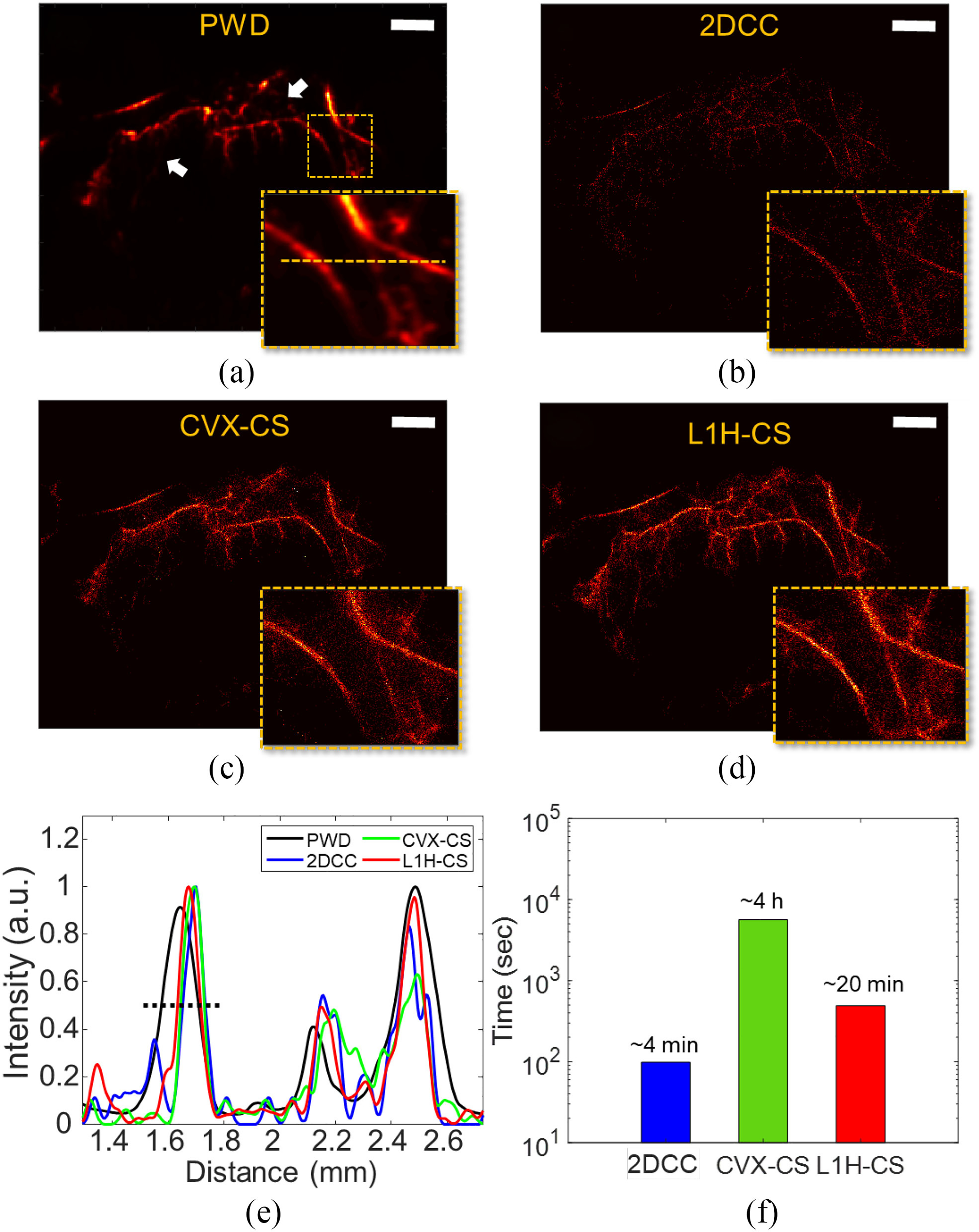
Fig. 5.

SRUS images of mouse tumor using (a) PWD, (b) 2DCC, (c) CVX-CS, and (d) L1H-CS algorithms, respectively. (e) Intensity profiles along the orange lines indicated in the inset image of (a). (f) Total computational time for the image size of 252 × 252 pixels with 500 frames. Scale bars indicate 1 mm.
