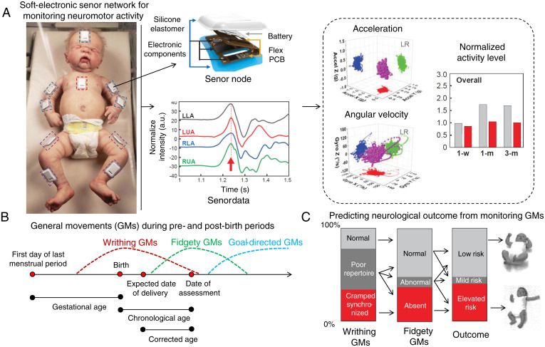
Fig. 1.
Soft-electronic sensor network for early detection of later neurological deficits in infants. (A) The sensors are placed on the forehead, chest, and limbs of the infants. These sensors are fabricated using flexible printed circuit boards (PCBs). Electronic components and batteries are assembled and encapsulated inside a waterproof silicone elastomer. Accelerometer and gyroscope data from the left upper arm (LUA), left lower arm (LLA), right upper arm (RUA), and right lower arm (RLA) are then interpreted to acceleration, angular velocity, and normalized activity levels. With machine-learning techniques, these three-axis accelerations and angular velocities are reconstructed to reveal typical/atypical GMs. Adapted from ref. 8. (B) GMs during pre- and postbirth periods. Three types of GMs: (1) writhing GMs, (2) fidgety GMs, and (3) goal-directed GMs are observed from early fetal life to 6 mo of age. Reprinted with permission from ref. 9. (C) Predicting neurological outcome from the occurrence of writhing and fidgety GMs. A longitudinal study on 130 infants showed that monitoring the writhing and fidgety GMs can be used to predict neurological outcome at 3 y of age. Adapted with permission from ref. 3.

