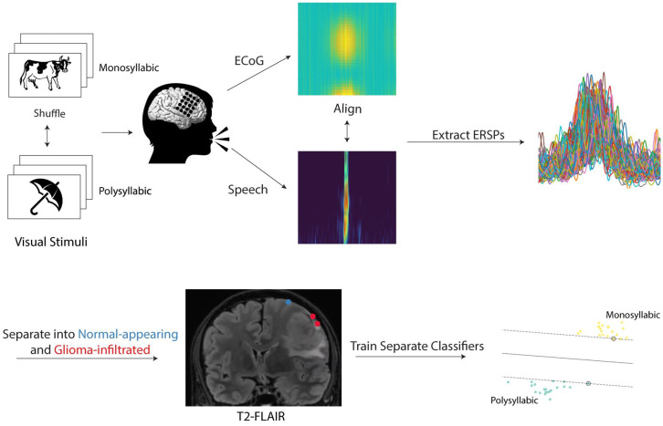
Fig. 1.
Experimental workflow. Participants with perisylvian gliomas on the surface of the brain were asked to complete a picture naming task during intraoperative functional mapping. On each trial, participants were presented with a line drawing of a common object or animal in a randomized fashion and prompted to provide its name. Of the 48 stimuli, 28 represented monosyllabic words while 20 represented polysyllabic words. Audio and electrophysiologic (via ECoG) recordings were taken from each participant during task completion and aligned to speech onset. ERSPs were extracted in the high-gamma range (70 to 170 Hz). Electrodes were localized on each participant’s preoperative T2-FLAIR image and categorized as “normal-appearing” if overlying normal-appearing cortex or “glioma-infiltrated” if overlying regions of T2-FLAIR signal abnormality. Using the ERSPs, separate classifiers were trained to decode the trial condition (monosyllabic versus polysyllabic) in normal-appearing and tumor-infiltrated cortex.

