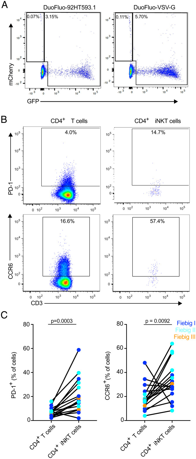
Fig. 7.
CD4+ iNKT cells express markers associated with the HIV reservoir. (A) Representative flow plots showing GFP and mCherry expression by iNKT cells 5 d postinfection with HIV-1 DuoFluo. Results are representative of three healthy donors. (B) Representative flow plots showing the expression of PD-1 and of CCR6 by peripheral blood and colonic CD4+ T cells and CD4+ iNKT cells. (C) Expression of markers enriched on latently infected cells, PD-1 (Left) and CCR6 (Right), by peripheral blood CD4+ T cells and CD4+ iNKT cells from ART-treated, HIV-infected individuals (n = 16).

