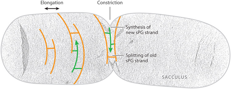
Figure 1.
Uranyl acetate–stained electron microscopy (EM) image of an isolated Escherichia coli sacculus with schematic drawing of the splitting of old (orange) and insertion of new (green) glycan strands in cell wall elongation and constriction. Figure adapted with permission from Reference 48. Abbreviation: sPG, septal peptidoglycan.

