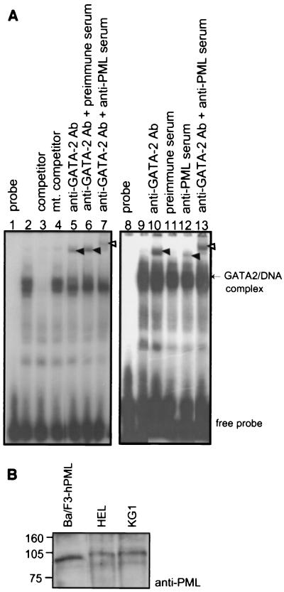
FIG. 4.
A GATA-2–PML complex binds to a GATA motif in DNA. (A) Nuclear extracts from Ba/F3-hPML cells were used in EMSAs. A 32P-labeled oligonucleotide containing GATA consensus recognition sites was used as a probe. The protein-DNA complex was revealed in lanes 2 and 9. Competitive experiments were performed using a 200-fold excess of either the unlabeled oligonucleotide (competitor) or a mutant (mt.) oligonucleotide in which the GATA consensus site had been disrupted (lanes 3 and 4). Supershift experiments were conducted by addition of anti-GATA-2 antibody (Ab; RC1.1) and/or anti-PML antiserum as indicated, with preimmune rabbit serum serving as a control (lanes 5 to 7 and 10 to 13). The positions of migration of free probe, specific GATA-2–DNA complexes, and the supershifted complexes (filled arrowheads) are indicated. The simultaneous addition of anti-PML and anti-GATA-2 antibodies to the reaction resulted in a further retarded complex indicated by the open arrowheads (lanes 7 and 13). (B) Nuclear extract from Ba/F3-hPML cells was analyzed for expression of hPML by Western blotting with anti-hPML antibody along with human hematopoietic HEL and KG1 cells. Equal numbers (106) of cells were used for the analysis. Sizes are indicated in kilodaltons.

