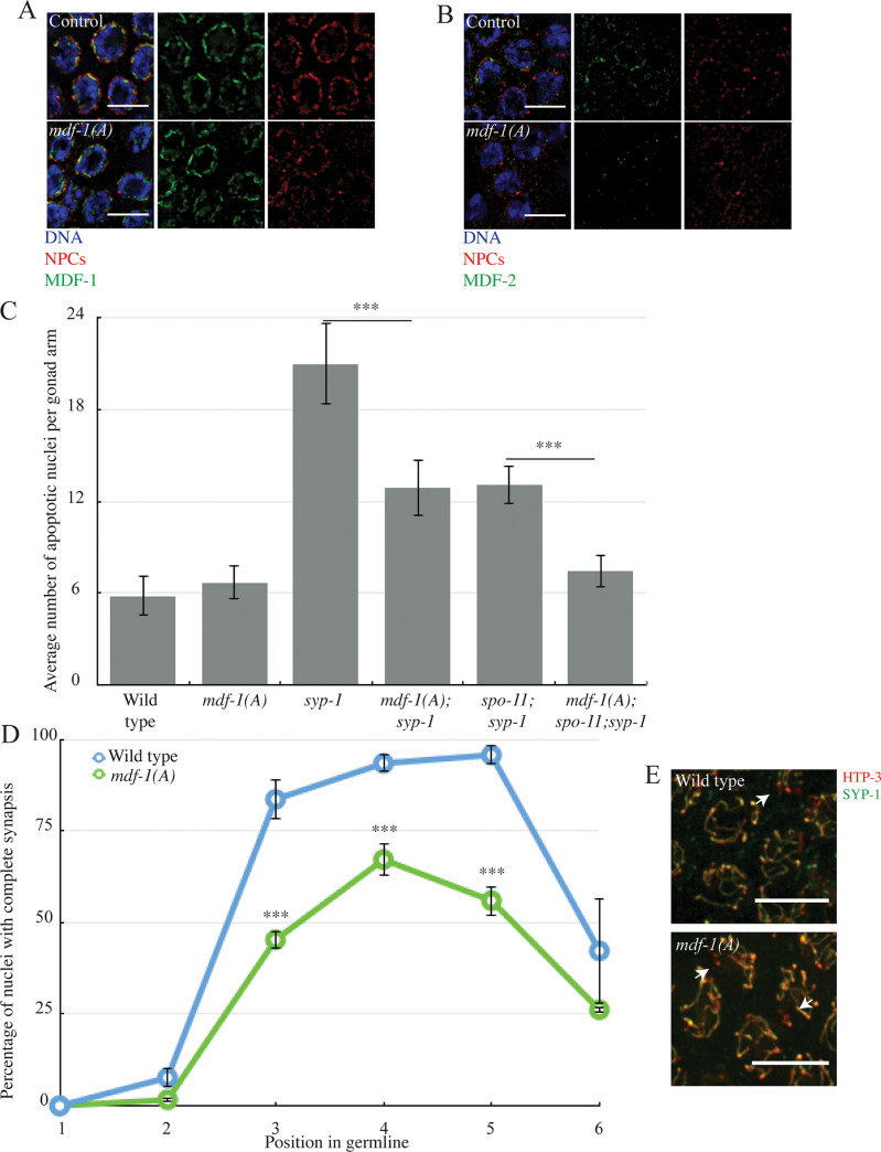
Fig 4. MDF-1MAD-1’s ability to interact with MDF-2MAD-2 is required to regulate and monitor synapsis.
A. MDF-1MAD-1 (A) localizes at the nuclear periphery. B. MDF-2MAD-2 (green) does not co-localize with NPCs (red) at the nuclear periphery in mdf-1mad-1(A) mutants. Images are partial projections of meiotic nuclei stained to visualize DNA (blue). Bar: 5 μm. C. mdf-1mad-1(A) reduces germline apoptosis in syp-1 and spo-11;syp-1 mutants. A *** indicates a p value < 0.0001. D. Synapsis is reduced and delayed in mdf-1mad-1(A) mutants. E. Images of nuclei during synapsis initiation in wild-type and mdf-1mad-1(A) mutants stained to visualize SYP-1 and HTP-3. Arrows indicates unsynapsed chromosomes. Bar: 5 μm.

