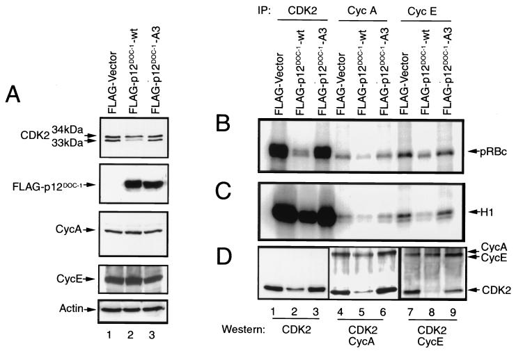
FIG. 5.
Ectopic expression of p12DOC-1 and CDK2 kinase activity in 293 cells. (A) Cellular levels of CDK2, FLAG-p12DOC-1-wt, FLAG-p12DOC-1-A3, cyclin A, cyclin E, and actin in control vector and p12DOC-1 transfectants. The samples were run on long SDS-PAGE gels to resolve the 33- and 34-kDa CDK2 bands. (B and C) In vitro phosphorylation using GST-pRBc and histone H1, respectively, as substrates. (D) CDK2, cyclin A, and cyclin E immunoblots to show intracellular levels of these proteins in p12DOC-1-wt (lanes 2, 5, and 8), p12DOC-1-A3 (lanes 3, 6, and 9), and control transfectants (lanes 1, 4, and 7). Lanes 1, 2, and 3, immunoblot for CDK2; lanes 4, 5, and 6, immunoblot for CDK2 and cyclin A; lanes 7, 8, and 9, immunoblot for CDK2 and cyclin E. Signals were quantified by exposing the probed membranes to a quantitative imaging system (Fluor-S MAX MultiImager; Bio-Rad). IP, immunoprecipitation.

