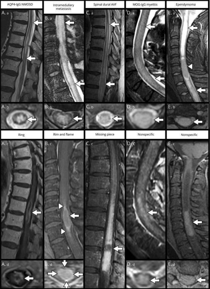
Figure 2. T2-Weighted and Postgadolinium T1-Weighted MRI Patterns in Various Myelopathy Diagnoses (Part 2).

A longitudinally extensive T2 hyperintense transverse myelitis (A.a-A.b, arrows) with associated elongated ring/partial ring enhancement (A.c-A.d, arrows) seen in AQP4-IgG NMOSD. A longitudinally extensive T2 hyperintense lesion (B.a-B.b, arrows) with an associated rim (arrows) and flame (arrowheads) pattern of enhancement surrounding a less enhancing center (B.c-B.d) seen in spinal cord intramedullary metastasis. Diffuse T2 hyperintensity (C.a-C.b, arrows) with apparent flow voids predominantly along the dorsal cord surface (C.a) and a missing piece of enhancement (C.c, arrow) in spinal dural arteriovenous fistula. Nonspecific T2 hyperintensity (D.a-D.b, arrows) and faint nonspecific contrast enhancement (D.c-D.d, arrows) seen in a patient with MOG-IgG associated myelitis. A T2 hyperintense expansile lesion (arrows) with mass effect (arrowhead) (E.a-E.b) and no clear pattern of associated contrast enhancement (E.c-E.d, arrows) seen in a patient with spinal cord ependymoma. (A) reprinted with permission from Sechi E, Flanagan EP. Autoimmune Demyelinating Syndromes: NMOSD and Anti-MOG Disease, Neuroimmunology, Springer, April 2021. (C) modified with permission from Zalewski NL, Rabinstein AA, Brinjikji W, et al. Unique Gadolinium Enhancement Pattern in Spinal Dural Arteriovenous Fistulas. JAMA Neurol 2018;75:1542-1545.
