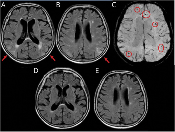
Figure 1. Spontaneous ARIA-like Imaging Findings at Presentation and After Radiologic Remission at 3-Month Follow-up of a Patient With Probable CAA-ri.
(A and B) Fluid-attenuated inversion recovery (FLAIR) images (axial sequences) at presentation show cortico-subcortical regions of hyperintensity suggestive of vasogenic edema and sulcal effacement on both hemispheres (red arrows). (C) Susceptibility-weighted images show microbleeds (red circles). (D and E) MRI 3 months after corticosteroid pulse therapy (5 IV boluses of 1 g/d methylprednisolone followed by 1 mg/kg oral prednisone daily and slow tapering off over several months) showing the disappearance of the acute inflammatory white matter hyperintensity lesions in the corresponding planes of FLAIR sequences indexed at presentation. ARIA = amyloid-related imaging abnormalities; CAA-ri = cerebral amyloid angiopathy–related inflammation.

