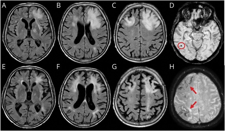
Figure 2. Spontaneous ARIA-like Imaging Findings of a Patient With Probable CAA-ri at Presentation and After 3 Months of Follow-up With No Radiologic Remission.
(A–C) Fluid-attenuated inversion recovery (FLAIR) axial sequences at presentation show cortico-subcortical hyperintense areas on both frontal lobes with edema and mass effect on the anterior horn of the left lateral ventricle. (E–G) FLAIR sequences 3 months after corticosteroid pulse therapy (5 IV boluses of 1 g/d methylprednisolone followed by 1 mg/kg oral prednisone daily and slow tapering off over several months) treatment showing substantial reduction of the signal abnormalities on the right frontal lobe and persistent hyperintense signal abnormality on the left frontal lobe without mass effect and with atrophy of the corresponding cortico-subcortical region. (D and H) Susceptibility-weighted imaging sequences at presentation show microbleeds (red circle) and diffuse cortical superficial siderosis (red arrows). ARIA = amyloid-related imaging abnormalities; CAA-ri = cerebral amyloid angiopathy–related inflammation.

