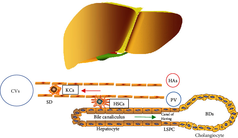
Figure 1.

The structure of liver tissue. Liver tissue accepts a dual blood supply from the portal vein (PV) and the hepatic artery (HA). Hepatocytes produce the bile, which flows into the bile ducts (BDs). Hepatic arterioles, terminal branches of the portal vein, and the most minor bile ducts form the compact portal tracts. Bile ductules drained into the bile ducts via the canals of Hering, which contain LSPCs. The narrow space between trabeculae and the endothelium of blood sinusoids is called the spaces of Disse (SD), which participate in the bidirectional traffic between blood and hepatocytes. In the case of acute injury or chronic liver diseases, hepatic progenitor cell activation occurs in response to different inflammatory cytokines. CVs: central veins; LSPCs: liver stem/progenitor cells; KCs: Kupffer cells; HSCs: hepatic stellate cells.
