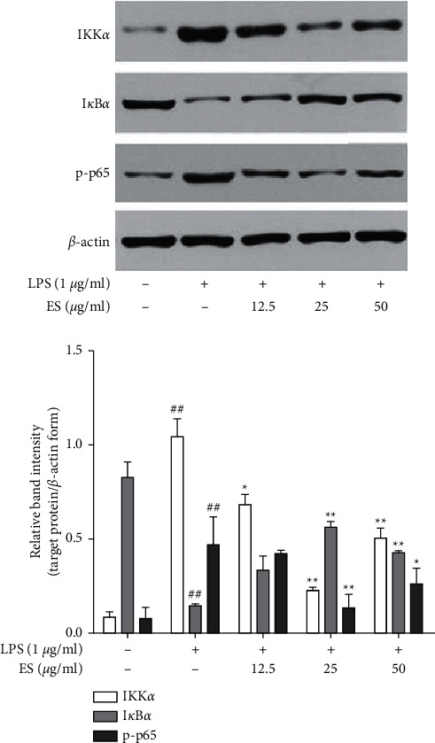
Figure 9.

Effect of ES on the NF-κB signaling pathway in LPS-induced THP-1 cells. Cells were treated with ES (12.5, 25, and 50 μg/mL) for 1 h then incubated with or without LPS (1 μg/mL) for 1 h. Protein expression was tested by Western blot (##P < 0.01 versus the blank control group; ∗P < 0.05 and ∗∗P < 0.01 versus the LPS-treated group).
