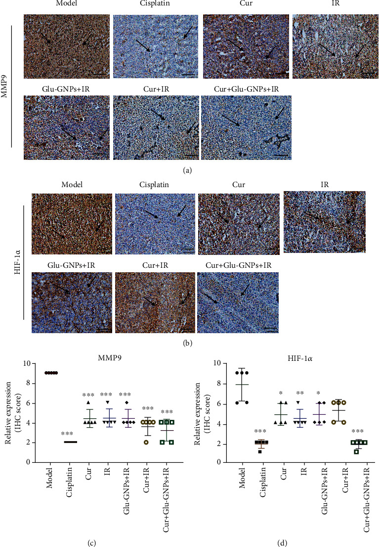
Figure 5.

The protein expressions of MMP9 and HIF-1α in tumor tissue samples by immunohistochemistry. (a) Representative images of MMP9 and (b) HIF-1α IHC staining (magnification, ×200). Tumor tissue was immunostained using DAB (brown) and hematoxylin (blue) for nuclear counterstaining. Arrows indicate the nuclear expression of MMP9 or HIF-1α in breast cancer cells (scale bars, 100 μm). (c) The semiquantitative scoring analysis of MMP9 and (d) HIF-1α protein expression in tumor tissue. IHC: immunohistochemistry; MMP9: matrix metalloprotein-9; HIF-1α: hypoxia-inducible factor-1α.
