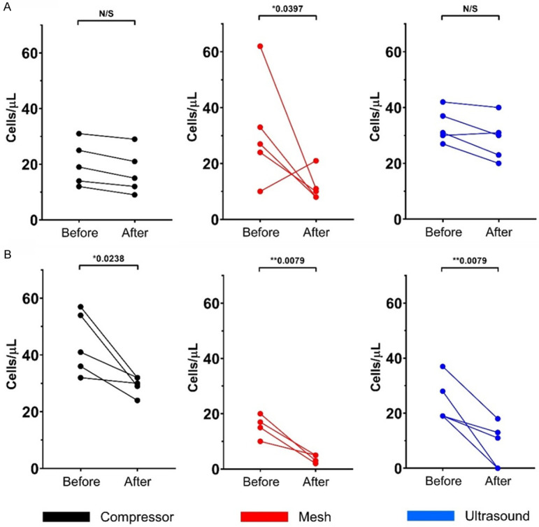
Figure 2.

Changes in nucleated absolute cells counts (cells/µL) analyzed by flow cytometry. Cells were stained with Hoechst 33342 before and after three nebulization methods; each color represents one nebulization method [black: compressor (n=5); red: mesh (n=5); and blue: ultrasound nebulization (n=5)]. A. CD45- nucleated cells. B. CD45dim nucleated cells. *Statistically significant differences (P<0.05). **Statistically highly significant differences (P<0.01).
